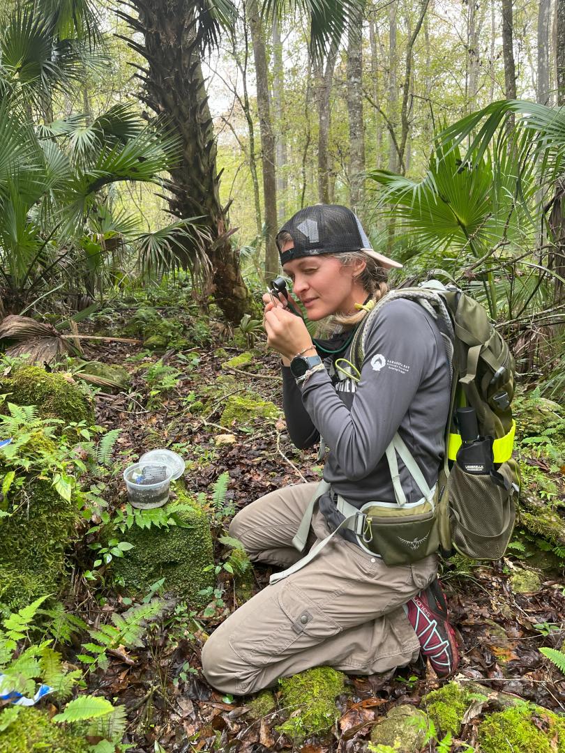
Kentucky is a botanically diverse state home to over 2,000 native plants and more than 400 taxa that are of conservation concern. Kentucky’s native plants are phylogenetically diverse, and a subset of taxa reflect Kentucky’s geologic history as tropical relicts. This is especially true for the ferns in Kentucky, as many species occupy sandstone rock shelters which buffer extreme climatic conditions much like cave ecosystems. These microclimatic pockets create unique distribution patterns for the ferns that occupy this niche space, and even further partition the fern life cycle such that some crevices host only gametophytes while others host both gametophyte and sporophyte generations. This talk will focus on decades of work conducted in these unique rock shelter environments and the spatial differentiation of fern generations (gametophyte/sporophyte) in Kentucky, the Appalachians and beyond. Ecological research focusing on topics such as local adaptation, physiological tolerance limits, and population differentiation will be discussed. The remainder of the talk will highlight the botanical resources housed at Eastern Kentucky University and the utility of these natural history collections to scientists worldwide.
Kentucky is a botanically diverse state home to over 2,000 native plants and more than 400 taxa that are of conservation concern. Kentucky’s native plants are phylogenetically diverse, and a subset of taxa reflect Kentucky’s geologic history as tropical relicts. This is especially true for the ferns in Kentucky, as many species occupy sandstone rock shelters which buffer extreme climatic conditions much like cave ecosystems. These microclimatic pockets create unique distribution patterns for the ferns that occupy this niche space, and even further partition the fern life cycle such that some crevices host only gametophytes while others host both gametophyte and sporophyte generations. This talk will focus on decades of work conducted in these unique rock shelter environments and the spatial differentiation of fern generations (gametophyte/sporophyte) in Kentucky, the Appalachians and beyond. Ecological research focusing on topics such as local adaptation, physiological tolerance limits, and population differentiation will be discussed. The remainder of the talk will highlight the botanical resources housed at Eastern Kentucky University and the utility of these natural history collections to scientists worldwide.
Kentucky is a botanically diverse state home to over 2,000 native plants and more than 400 taxa that are of conservation concern. Kentucky’s native plants are phylogenetically diverse, and a subset of taxa reflect Kentucky’s geologic history as tropical relicts. This is especially true for the ferns in Kentucky, as many species occupy sandstone rock shelters which buffer extreme climatic conditions much like cave ecosystems. These microclimatic pockets create unique distribution patterns for the ferns that occupy this niche space, and even further partition the fern life cycle such that some crevices host only gametophytes while others host both gametophyte and sporophyte generations. This talk will focus on decades of work conducted in these unique rock shelter environments and the spatial differentiation of fern generations (gametophyte/sporophyte) in Kentucky, the Appalachians and beyond. Ecological research focusing on topics such as local adaptation, physiological tolerance limits, and population differentiation will be discussed. The remainder of the talk will highlight the botanical resources housed at Eastern Kentucky University and the utility of these natural history collections to scientists worldwide.
The cerebral cortex is arguably the brain area that underwent the most profound transformations in vertebrate brain evolution. The expansion of the cerebral cortex in mammals was accompanied by an explosion of neuronal diversity. To discover general principles underlying the evolution of neuron types and circuits, we study the simple cerebral cortices of non-mammalian vertebrates. Our recent work has focused on the Spanish newt Pleurodeles waltl, a species with a key phylogenetic position in the vertebrate tree. We are investigating the neuroanatomy, cell type composition, and function of the Pleurodeles brain using a combination of modern neuroscience tools.
Our work on amphibians and reptiles indicates that the cerebral cortex of ancestral tetrapods was layered, with two main classes of neurons with distinct laminar positions, molecular identities, and long-range projections. In salamanders, these two layers are generated sequentially from multipotent progenitors in an outside-in sequence. We propose that in mammals new types of pyramidal neurons evolved from these two ancestral classes by diversification, through the emergence of novel gene regulatory interactions during neuronal differentiation.
Dr. Samantha Brugmann is a developmental biologist studying craniofacial development and disease. Her longterm goal is to help children with craniofacial anomalies by generating tissue amenable for surgical repair. To achieve this goal, her lab specifically focuses on the role the primary cilium during craniofacial development and the craniofacial anomalies that arise when the cilium do not function properly. Projects in her lab utilize avian, murine and humaninduced pluripotent stem cells to gain a better understanding of the molecular mechanisms associated with craniofacial anomalies. In addition to using existing animal models to understand human craniofacial disorders, her lab also sequences patients and generates cell-based models to uncover novel genetic causes for craniofacial ciliopathies.
Abstract:
The Brugmann Lab focuses on the understanding molecular and cellular processes important for craniofacial development and the onset of craniofacial anomalies (CFAs). CFAs represent approximately one third of all birth-defects. For the past decade, my research program has centered on treating these conditions by garnering a fundamental understanding of craniofacial development and pathological mechanisms associated with CFAs. We have specifically focused on a class of CFAs called ciliopathies, which are caused by disruptions to a cellular organelle called the primary cilium. Ciliopathies represent a fast-growing group of disorders, that can affect up to 1 in 800 people. My lab was the first to report that the craniofacial complex is the primary organ system affected in 30% of all ciliopathies, and thus coined the term craniofacial ciliopathies. My lab uses murine, avian and human model systems to understand molecular mechanisms associated with ciliopathies. Furthermore, we use these model systems to identify potential therapeutic avenues to treat this class of diseases.

 Dr. Sally Chambers